X-ray CT Imaging
Computed tomography: principles and image reconstruction
About This Series
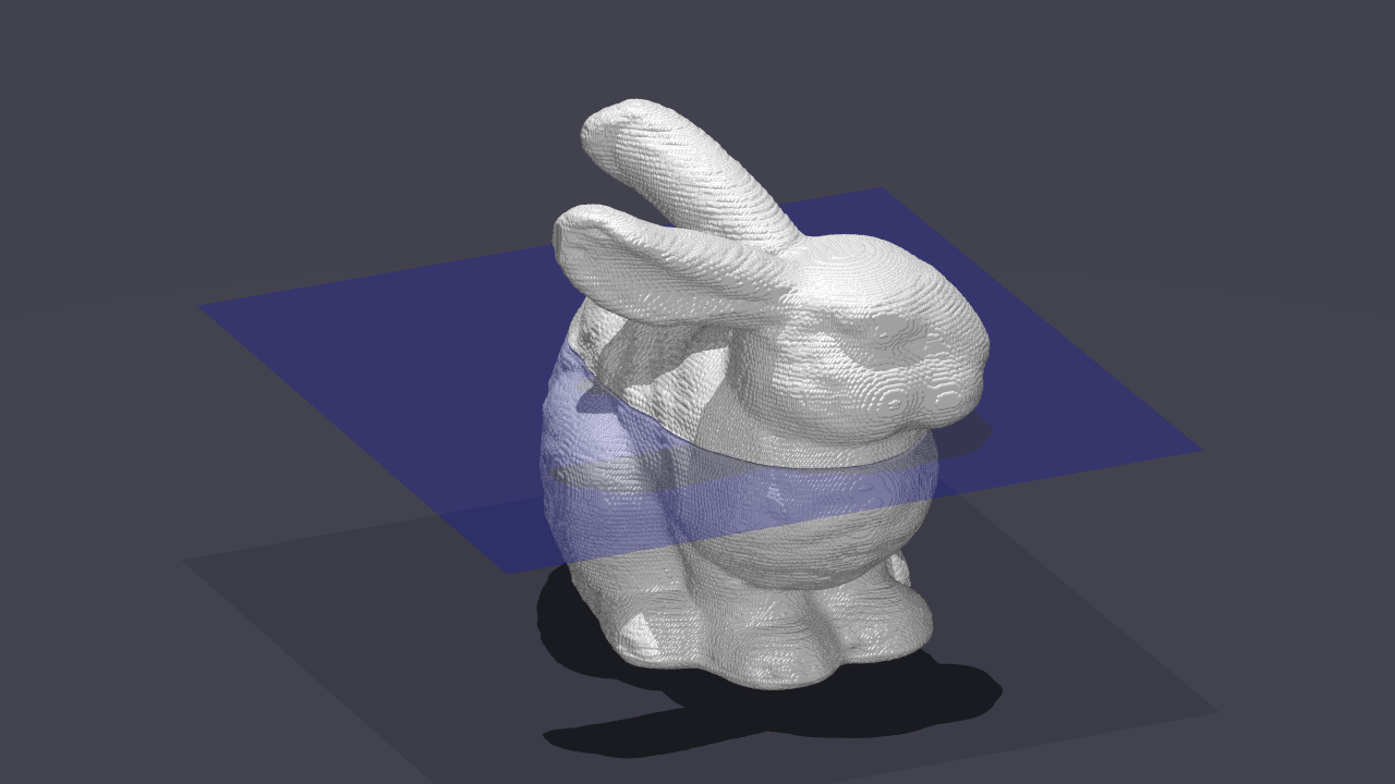
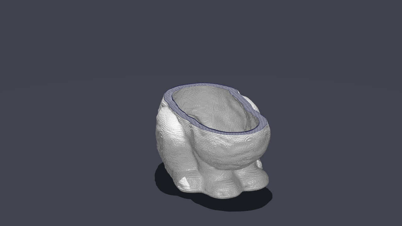
X-ray CT (computed tomography) is a technology for visualizing the internal structure of an object nondestructively using X-rays. It is used across a wide range of fields, including medical diagnosis, industrial inspection, and materials science.
This series proceeds step by step from the physics of X-rays through the mathematical formulation via the Radon transform, image reconstruction by filtered back-projection (FBP), and on to the latest iterative and deep-learning approaches.
The fundamental equation of X-ray CT
$$I = I_0 \exp\left(-\int_L \mu(x, y) \, dl\right)$$$\mu(x, y)$: linear attenuation coefficient; $L$: X-ray path
Taking the logarithm of both sides gives $-\log(I/I_0) = \int_L \mu\,dl$, the line integral of the attenuation coefficient $\mu$ along the path — that is, the Radon transform itself (assuming monochromatic X-rays; real X-rays are polychromatic, which causes beam-hardening artifacts). CT reconstruction therefore reduces to the inverse problem of recovering $\mu(x, y)$ from projection data taken from many directions.
Learn by Level
Learning Path
Application Areas
Medical diagnosis
Detecting and diagnosing disease with head, chest, and abdominal CT. A wide variety of protocols exist, such as coronary CT and contrast-enhanced CT.
Industrial inspection
Nondestructive inspection of welds, detection of internal defects in castings, and inspection of solder joints on electronic boards.
Security
Automatic detection of dangerous goods in airport baggage screening. Material identification with dual-energy CT.
Cultural heritage and archaeology
Analyzing the internal structure of mummies and ancient artifacts, investigating their contents nondestructively.
Key Concepts and Formulas
Beer–Lambert law
$$I = I_0 \exp\!\left(-\int_L \mu(x,y)\,dl\right)$$
Radon transform
$$\mathcal{R}f(s,\theta) = \int_{-\infty}^{\infty}\!\int_{-\infty}^{\infty} f(x,y)\,\delta(x\cos\theta + y\sin\theta - s)\,dx\,dy$$
Fourier slice theorem
$$P(\omega,\theta) = F(\omega\cos\theta,\;\omega\sin\theta)$$
Filtered back-projection (FBP)
$$f(x,y) = \int_0^{\pi}\! \left[\mathcal{R}f(s,\theta) * h(s)\right]_{s=x\cos\theta+y\sin\theta} d\theta$$
Historical Background
The principle of X-ray CT goes back to 1917, when Johann Radon established the mathematical theory of the Radon transform. The first practical CT scanner was developed in 1971 by Godfrey Hounsfield (EMI), and the South African physicist Allan Cormack had independently published the reconstruction theory earlier (1963 and 1964). The two shared the Nobel Prize in Physiology or Medicine in 1979.
Today the technology continues to advance rapidly, with multi-slice CT, dual-energy CT, photon-counting CT, and more.
Prerequisites
- Introductory: basic knowledge of X-rays (middle-school science), basics of trigonometry
- Basic: basics of linear algebra, calculus (integration)
- Intermediate: the Fourier transform, basics of signal processing, linear algebra (eigenvalues, matrix decomposition)
- Advanced: optimization theory, statistical estimation, basics of deep learning
Related Topics
- Radon transform - the mathematical foundation of CT reconstruction
- Fourier analysis - the Fourier slice theorem
- Filter design - choosing the filter for FBP
- Optimization - the basis of iterative reconstruction (ART, MBIR)
- Linear algebra - formulating the projection operator and the inverse problem
Frequently Asked Questions
What is X-ray CT?
X-ray CT (computed tomography) is a nondestructive inspection technology that mathematically reconstructs cross-sectional images of the interior of an object from projection data obtained by irradiating it with X-rays from many directions.
What are the main methods of CT image reconstruction?
Representative methods include filtered back-projection (FBP), algebraic iterative reconstruction (ART), statistical reconstruction (MBIR), and deep-learning-based methods. FBP has long been the standard (iterative reconstruction has also become widespread in recent clinical CT).
What is the Fourier slice theorem?
It is the theorem that the one-dimensional Fourier transform of a parallel-beam projection equals the values along the corresponding slice (a straight line) of the two-dimensional Fourier transform of the original image. It is the mathematical foundation of CT image reconstruction.
Why does CT need to image from many directions?
A projection from a single direction cannot tell where along the X-ray path the attenuation occurred, so the internal structure cannot be determined uniquely. Collecting projections from a sufficient number and angular range of directions (too few directions or a limited angular range is not enough) provides enough information to recover the attenuation coefficient at each point and reconstruct a cross-sectional image. Mathematically, this corresponds to solving the inverse problem of the Radon transform.